Breast cancer screening has become more accessible and sophisticated, but the process doesn’t end with a mammogram. When imaging reveals an abnormality—a mass, calcifications, or architectural distortion—the next step often involves obtaining a tissue sample to determine whether the finding is benign or malignant. This diagnostic phase can be unsettling, yet understanding what happens when screening detects something unusual helps patients approach the process with realistic expectations rather than fear.
Why Imaging Alone Cannot Provide a Definitive Answer
Mammograms, ultrasounds, and MRI scans excel at detecting structural changes in breast tissue, but they cannot differentiate between cancer and benign conditions with absolute certainty. A solid mass might be a fibroadenoma, a cyst with thick walls, or a malignant tumor. Calcifications could represent harmless calcium deposits or early-stage ductal carcinoma in situ. Radiologists assign suspicion levels based on imaging characteristics, but tissue analysis remains the diagnostic standard.
The Breast Imaging Reporting and Data System (BI-RADS) categorizes findings from 0 to 6, with categories 4 and 5 indicating sufficient concern to warrant biopsy. Category 4 findings have a 2 to 95 percent likelihood of malignancy depending on subcategory, while category 5 findings show a 95 percent or higher probability. These percentages underscore why visual assessment alone cannot replace cellular examination.
How Tissue Sampling Works in Modern Breast Diagnosis
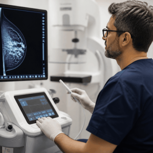
When biopsy becomes necessary, the approach depends on what imaging modality best visualizes the abnormality. Ultrasound-guided procedures work well for masses visible on sonography, offering real-time needle placement without radiation. Stereotactic biopsy uses mammographic guidance for calcifications or distortions seen only on X-ray, while MRI-guided techniques target lesions visible exclusively on magnetic resonance imaging.
Core needle biopsy has largely replaced surgical excision as the initial diagnostic method. Using a spring-loaded device, the radiologist obtains multiple tissue cores—typically three to six samples—through a single insertion point. The procedure takes 20 to 30 minutes, performed under local anesthesia, and allows patients to resume normal activities within a day. A small marker clip placed at the biopsy site ensures the abnormality can be located later if treatment becomes necessary.
The Role of Vacuum-Assisted Devices
For certain findings, vacuum-assisted biopsy provides advantages over standard core technique. The device removes larger, contiguous tissue samples through a single needle insertion, which proves particularly useful for sampling calcifications or when complete removal of a small lesion is desirable. Some benign findings, such as papillomas or radial scars, can be excised entirely through this minimally invasive approach, potentially eliminating the need for surgical follow-up.
What Happens After Tissue Collection
Biopsy samples undergo processing that takes several days. A pathologist examines the tissue under microscopy, looking for cellular abnormalities, growth patterns, and architectural changes that distinguish normal tissue from precancerous conditions or invasive cancer. If malignancy is confirmed, additional tests determine hormone receptor status, HER2 expression, and proliferation rate—information that directly influences treatment selection.
Results typically arrive within three to five business days, though complex cases requiring additional staining or expert consultation may take longer. Approximately 20 to 30 percent of biopsies performed for suspicious findings reveal cancer, meaning the majority return benign results. Even when cancer is diagnosed, early detection through screening generally means smaller tumors and more treatment options.
Accessing Specialized Diagnostic Services
Comprehensive breast programs integrate imaging and biopsy services with multidisciplinary care teams, streamlining the diagnostic process and reducing the time between detection and definitive answers. For women in the United Arab Emirates, Cleveland Clinic Abu Dhabi’s Breast Health & Cancer Program offers the full spectrum of diagnostic capabilities, including image-guided breast biopsy procedure options performed by specialized radiologists. The facility’s multidisciplinary approach means that if biopsy results require treatment, patients can immediately access medical oncology, surgical expertise, and reconstruction services without navigating multiple healthcare systems.
Managing Anxiety During the Diagnostic Window
The period between biopsy and results represents an unavoidable waiting phase that many patients find challenging. While healthcare providers cannot accelerate tissue processing, they can offer realistic timeframes and explain what the pathology examination entails. Understanding that most biopsies return benign results provides statistical reassurance, though it does not eliminate individual concern.
Practical strategies include maintaining normal routines rather than putting life on hold, limiting repetitive internet searches that often increase rather than reduce anxiety, and identifying one or two trusted information sources rather than consulting multiple conflicting websites. Some patients find it helpful to prepare questions for the results appointment in advance, ensuring they address their concerns regardless of emotional state when news arrives.
Privacy Considerations in Healthcare Data
Diagnostic procedures generate sensitive health information that requires protection under privacy regulations. Patients should understand how their biopsy results, imaging studies, and related medical records are stored, shared, and secured. Healthcare providers follow strict protocols regarding who can access this information and under what circumstances. For those concerned about broader data privacy practices, resources like consumer privacy rights information can provide context about how personal information is handled across different sectors, though healthcare data faces additional regulatory oversight through specific medical privacy laws.
Evolving Screening Recommendations and Their Impact
Recent changes to breast cancer screening guidelines reflect accumulating evidence about benefits and harms across different age groups. The U.S. Preventive Services Task Force now recommends that women begin biennial mammography at age 40 rather than 50, a shift prompted by rising breast cancer rates among younger women and refined understanding of screening effectiveness. These updated mammogram screening recommendations aim to detect more cancers at earlier stages when treatment is most effective, though they also increase the number of callbacks and biopsies performed on benign findings.
The guideline revision highlights the tension inherent in screening programs: casting a wider net catches more significant disease but also generates more false alarms. Women beginning screening at 40 will likely experience more callbacks and biopsies over their lifetime compared to those who start at 50, yet they will also benefit from earlier detection of real cancers. Understanding this trade-off helps contextualize the biopsy experience as a feature of effective screening rather than an indication that something has gone wrong.
Moving Forward After Diagnosis
Whether biopsy results reveal cancer or confirm benign findings, the experience often shifts a patient’s relationship with breast health. Those receiving benign diagnoses typically return to routine screening schedules, though some findings may warrant shorter intervals initially. Women diagnosed with high-risk lesions such as atypical hyperplasia may enter enhanced surveillance programs or consider risk-reduction strategies. Cancer diagnoses lead to treatment planning discussions that address surgery, radiation, systemic therapy, and reconstruction based on tumor characteristics and patient preferences.
The diagnostic pathway from screening abnormality to definitive tissue diagnosis represents a necessary bridge between detection and appropriate action. While the process involves uncertainty and waiting, modern biopsy techniques minimize discomfort and provide the cellular information required for sound medical decision-making. Understanding what happens during this interval—and why it happens—helps patients approach breast health with informed perspective rather than ungrounded fear.